Ranchi College of Pharmacy, Ranchi, Jharkhand 834003, India
The nervous system represents one of the most intricate and dynamic networks in living organisms, orchestrating a vast array of physiological processes, behavioral responses, and cognitive functions essential for survival and adaptation. This review delves into the multifaceted organization and operation of the nervous system, providing an in-depth examination of its structural framework, cellular composition, and underlying physiological mechanisms. Key components such as the central nervous system (CNS) and peripheral nervous system (PNS) are explored, with a particular focus on their roles in sensory perception, motor coordination, and homeostatic regulation. The interplay between neurons and glial cells, including astrocytes, oligodendrocytes, and microglia, is highlighted, emphasizing their collective contribution to neural signaling and maintenance. Advances in neuroscience have expanded our understanding of neural plasticity—the capacity of the nervous system to adapt to environmental changes and injury—and the molecular pathways governing synaptic transmission and regeneration. Genetic and environmental influences on neural development and function are also critically assessed, shedding light on their role in both normal physiology and pathophysiology. Furthermore, this review addresses a spectrum of nervous system disorders, from neurodegenerative diseases like Alzheimer’s and Parkinson’s to psychiatric conditions such as depression and schizophrenia, underscoring their complex etiologies and clinical manifestations. Emerging therapeutic strategies, including stem cell therapy, neuroprosthetics, and advanced neuroimaging techniques, are discussed in the context of their potential to revolutionize treatment paradigms and improve patient outcomes. This synthesis of current research and translational applications provides a comprehensive perspective on the nervous system's pivotal functions and its centrality to health and disease.
The nervous system represents a sophisticated biological network that fundamentally defines the operational capabilities of multicellular organisms. This intricate arrangement of specialized cells and tissues operates through precisely coordinated electrical and chemical signaling mechanisms to maintain homeostasis and facilitate organism-environment interactions. At its core, the nervous system comprises two main divisions: the central nervous system (CNS), encompassing the brain and spinal cord, and the peripheral nervous system (PNS), which includes cranial and spinal nerves extending throughout the body. The cellular foundation of neural function lies in neurons, highly specialized cells characterized by their distinctive morphology and electrochemical properties. These cells possess elaborate dendritic arbors for receiving inputs, a cell body containing essential molecular machinery, and axons capable of rapid signal propagation. Supporting these neurons are glial cells, including astrocytes, oligodendrocytes, and microglia in the CNS, and Schwann cells in the PNS, which maintain optimal conditions for neural function through various mechanisms such as myelination, metabolic support, and immune defence. Signal transmission within the nervous system occurs through both electrical and chemical mechanisms. Action pote ntials, generated through the coordinated activity of voltage-gated ion channels, propagate along axons as electrical signals. At synapses, these electrical signals trigger the release of neurotransmitters, which bind to specific receptors on target cells, thereby converting electrical signals into chemical messages that can either excite or inhibit subsequent neural activity. The functional organization of the nervous system exhibits remarkable hierarchical complexity. At the molecular level, neurotransmitter systems, including acetylcholine, dopamine, serotonin, and various neuropeptides, regulate specific aspects of neural function. These systems interact within neural circuits that process specific types of information, such as visual perception, motor control, or emotional responses. These circuits, in turn, are integrated into larger networks that coordinate complex behaviors and cognitive processes. The nervous system demonstrates remarkable plasticity, allowing for adaptation to environmental changes and learning from experience. This adaptability operates through multiple mechanisms, including synaptic modification, dendritic remodeling, and the integration of newly generated neurons in specific brain regions. Such plasticity underlies essential processes like memory formation, skill acquisition, and recovery from injury. Recent technological advances have revolutionized our understanding of nervous system function. Techniques such as optogenetics, which allows for precise control of specific neural populations using light, and advanced imaging methods that enable visualization of neural activity in real-time, have provided unprecedented insights into neural circuit operation. Additionally, the emergence of sophisticated computational models has enhanced our ability to understand how neural networks process information and generate behaviour. Dysfunction of the nervous system underlies numerous pathological conditions, ranging from neurodevelopmental disorders to neurodegenerative diseases. Understanding the fundamental principles of nervous system organization and function has crucial implications for developing therapeutic strategies for these conditions. Current research focuses on various approaches, including stem cell therapy, gene editing, and development of novel pharmacological agents targeting specific neural circuits or molecular pathways. This intricate biological system continues to be a frontier of scientific investigation, with new discoveries regularly emerging about its organization and function. As our understanding deepens, particularly through the application of emerging technologies and analytical approaches, we gain increasingly sophisticated insights into how the nervous system enables organisms to perceive, respond to, and interact with their environment while maintaining internal homeostasis and supporting complex cognitive processes [1, 2].
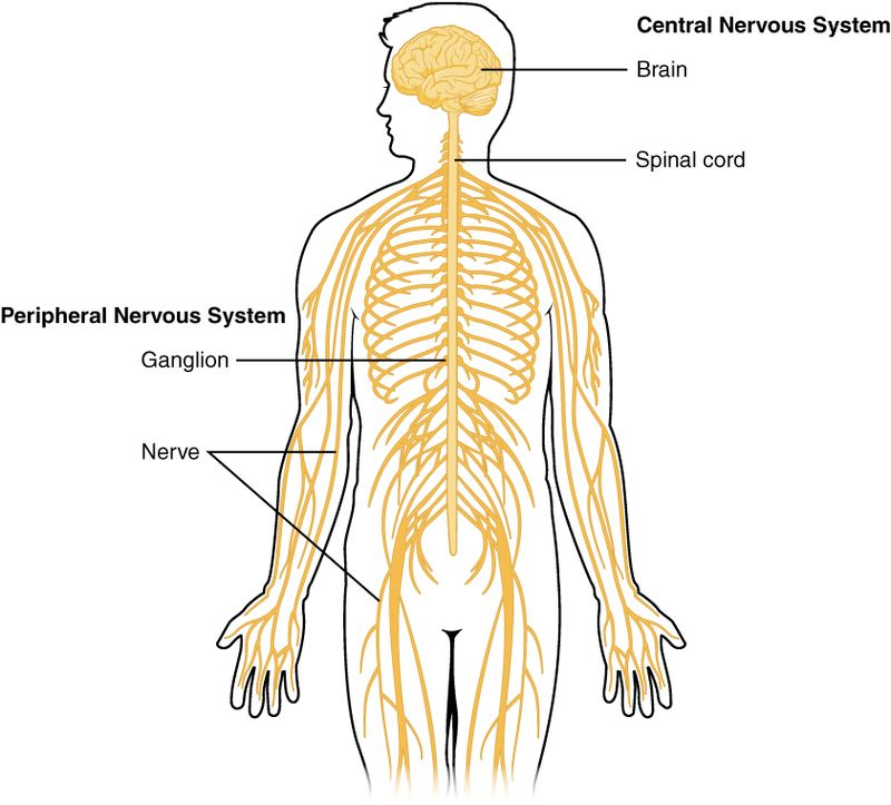
Fig. 1. Nervous System
The nervous system is divided into two major components: the central nervous system (CNS) and the peripheral nervous system (PNS). Each plays a distinct yet interconnected role in maintaining physiological and behavioral functions.
The central nervous system (CNS) represents the primary control center of the body, consisting primarily of the brain and spinal cord. This sophisticated network processes and integrates information from throughout the body, coordinating responses and maintaining homeostasis.
The CNS is encased within protective structures: the skull (cranium) houses the brain, while the vertebral column protects the spinal cord. Three layers of meninges – the dura mater, arachnoid mater, and pia mater – provide additional protection and support. Between the arachnoid and pia mater lies the subarachnoid space, filled with cerebrospinal fluid (CSF), which acts as a shock absorber and helps maintain optimal chemical conditions for neural function.
The brain, weighing approximately 1.4 kilograms in adults, consists of several major regions:
The cerebrum, the largest portion, is divided into two hemispheres connected by the corpus callosum. Its outer layer, the cerebral cortex, contains six distinct layers of neurons and is organized into four main lobes:
The diencephalon, located beneath the cerebrum, contains the thalamus (sensory relay station) and hypothalamus (homeostatic control center). The brainstem, comprising the midbrain, pons, and medulla oblongata, controls vital functions like breathing, heart rate, and blood pressure.
The cerebellum, positioned posterior to the brainstem, coordinates movement, balance, and motor learning. Its highly folded surface contains more neurons than the rest of the brain combined, despite occupying only 10% of total brain volume.
The spinal cord, a crucial component of the central nervous system, extends caudally from the medulla oblongata to the lumbar region. Functioning as a vital conduit, it houses neural pathways that facilitate communication between the brain and the peripheral nervous system. Upon cross-sectional examination, the spinal cord exhibits a distinct structural organization. Gray matter, characterized by the presence of neuronal cell bodies, occupies the central region, often described as a butterfly-shaped configuration. Surrounding the gray matter lies the white matter, composed of densely packed myelinated axons. These axons are meticulously organized into ascending and descending tracts, responsible for transmitting sensory and motor information, respectively. Furthermore, a central canal traverses the longitudinal axis of the spinal cord, containing cerebrospinal fluid (CSF) which provides vital support and protection to the delicate neural tissues.
The CNS comprises two main cell types:
The CNS operates through various integrated systems:
This comprehensive blood supply network is vital for the CNS to meet its high metabolic demands and sustain its critical functions, including sensory perception, motor control, and cognitive processes. The blood-brain barrier strictly regulates substance exchange between blood and neural tissue, maintaining a stable environment for optimal neural function.
The complex organization and function of the CNS enable sophisticated information processing and behavioral control, making it essential for survival and higher cognitive functions. Ongoing research continues to reveal new aspects of CNS function and potential therapeutic approaches for neurological disorders.
This intricate system's proper functioning is crucial for maintaining health and well-being, while its dysfunction can lead to various neurological and psychiatric conditions. Understanding its structure and function remains a central focus of neuroscience research and medical practice [3-7].
The Peripheral Nervous System (PNS) functions as a complex network of neural tissues that establishes vital connections between the central nervous system and various body structures. This sophisticated system comprises nerves, ganglia, and specialized receptors that extend throughout the organism, facilitating bidirectional information flow between central processing centers and peripheral targets.
Structurally, the PNS consists of two major functional divisions: the somatic nervous system and the autonomic nervous system. The somatic component primarily governs voluntary muscular activities and processes sensory information from the external environment. It encompasses both afferent pathways, which transmit sensory data from peripheral receptors to the CNS, and efferent pathways, which relay motor commands from the CNS to skeletal muscles. These neural circuits enable precise control of voluntary movements and facilitate rapid responses to environmental stimuli. The autonomic nervous system, conversely, regulates involuntary physiological processes and maintains homeostatic balance. This system further subdivides into sympathetic and parasympathetic branches, which generally exert opposing effects on target organs. The sympathetic division, often associated with "fight-or-flight" responses, utilizes norepinephrine as its primary neurotransmitter and increases physiological arousal. It accelerates heart rate, dilates bronchioles, inhibits digestive processes, and mobilizes energy reserves. The parasympathetic division, mediated primarily by acetylcholine, promotes "rest-and-digest" functions, reducing heart rate, facilitating digestion, and conserving energy.
At the cellular level, PNS neurons exhibit distinct structural and functional characteristics. Their cell bodies cluster in ganglia located outside the CNS, while their axons, often bundled into nerves, extend considerable distances to reach target tissues. These neurons utilize various neurotransmitters and neuromodulators to communicate with effector organs and other neural elements. Schwann cells provide myelin insulation for PNS axons, enhancing signal conduction velocity and maintaining axonal integrity. The PNS also contains specialized sensory receptors that detect specific environmental stimuli. These include mechanoreceptors for touch and pressure, thermoreceptors for temperature, nociceptors for pain, and proprioceptors for body position. These receptors transduce physical or chemical stimuli into electrical signals, which travel along sensory neurons to the CNS for processing. PNS function relies heavily on precise molecular mechanisms governing axonal transport, neurotransmitter synthesis and release, and receptor-mediated signaling. These processes ensure efficient communication between the CNS and peripheral tissues, enabling rapid and appropriate responses to both internal and external changes. Additionally, the PNS demonstrates remarkable regenerative capacity following injury, unlike the CNS, due to the supportive environment provided by Schwann cells and the expression of growth-promoting factors.
Understanding PNS organization and function proves crucial for medical practice, particularly in diagnosing and treating peripheral neuropathies, autonomic disorders, and neuromuscular conditions. Recent research continues to reveal new aspects of PNS function, including its role in neuroimmune interactions and its potential for therapeutic intervention in various pathological conditions [8-10].
Table 1. Overview of the Structural Organization of the Nervous System
|
Component |
Subcomponent |
Description |
|
Central Nervous System (CNS) |
Anatomical Organization |
Comprises the brain and spinal cord, protected by the skull, vertebral column, meninges, and cerebrospinal fluid (CSF). |
|
Brain Structure and Components |
Includes the cerebrum (divided into lobes: frontal, parietal, temporal, occipital), diencephalon (thalamus, hypothalamus), brainstem (midbrain, pons, medulla), and cerebellum. |
|
|
Spinal Cord Organization |
Features a butterfly-shaped gray matter core surrounded by white matter, organized into ascending (sensory) and descending (motor) tracts. |
|
|
Neural Tissue Components |
Composed of neurons (signal transmitters) and glial cells (support and maintenance), including astrocytes, oligodendrocytes, microglia, and ependymal cells. |
|
|
Functional Systems |
Sensory, motor, integrative, and communication systems for perception, voluntary movement, and autonomic regulation. |
|
|
Blood Supply and Metabolism |
Supported by the internal carotid arteries, vertebral arteries, and Circle of Willis; protected by the blood-brain barrier. |
|
|
Protection Mechanisms |
Includes physical protection (skull, vertebrae, meninges, CSF) and chemical barriers (blood-brain barrier, immune response). |
|
|
Development and Plasticity |
CNS exhibits neuroplasticity for learning, adaptation, and recovery from injuries. |
|
|
Communication Systems |
Utilizes electrical action potentials, neurotransmitters, and gap junctions for rapid and precise signaling. |
|
|
Peripheral Nervous System (PNS) |
Somatic Nervous System |
Controls voluntary muscular activities; includes afferent (sensory) and efferent (motor) pathways for communication with skeletal muscles. |
|
Autonomic Nervous System |
Regulates involuntary functions; divided into sympathetic ("fight-or-flight") and parasympathetic ("rest-and-digest") divisions. |
|
|
Neuronal Organization |
Neurons in ganglia outside the CNS; Schwann cells provide myelin for enhanced signal conduction. |
|
|
Sensory Receptors |
Specialized receptors (e.g., mechanoreceptors, thermoreceptors, nociceptors) transduce environmental stimuli into electrical signals for CNS processing. |
|
|
Regenerative Capacity |
Exhibits high regenerative ability due to Schwann cells and growth-promoting factors, unlike the CNS. |

Fig. 2. Human Brain

Fig. 3. Human Spinal Cord
The nervous system consists of two primary cell types: neurons and glial cells.
Neurons represent the fundamental cellular units of the nervous system, orchestrating the transmission of information through both electrical and chemical signaling mechanisms. These specialized cells exhibit distinct morphological and functional characteristics that enable complex neural processing.
The neuronal architecture comprises three primary components. The cell body, or soma, houses the nucleus and essential organelles responsible for protein synthesis and cellular maintenance. Dendrites extend from the soma like branching trees, creating elaborate networks that receive incoming signals from neighboring neurons. The axon, a specialized projection, conducts electrical impulses away from the soma toward target cells, often extending considerable distances through neural tissue. Each neuron possesses unique structural modifications that optimize its function. The axon terminal contains synaptic vesicles filled with neurotransmitters, while many axons feature myelin sheaths that enhance signal conduction velocity. Dendritic spines increase the surface area available for synaptic connections, facilitating robust neural networks.
Neurons can be categorized into three main functional classes based on their roles in neural circuits: Sensory neurons, also known as afferent neurons, specialize in detecting environmental stimuli and converting them into electrical signals. These neurons feature specialized receptor endings that respond to specific stimuli such as pressure, temperature, or chemical changes. They transmit this information from the periphery to the central nervous system for processing.
Motor neurons, or efferent neurons, conduct commands from the central nervous system to effector organs, primarily muscles and glands. These neurons typically have long axons and forms specialized junctions called motor end plates with muscle fibers, enabling precise control of movement and secretion.
Interneurons serve as intermediate processors within neural circuits, forming complex networks that integrate information between sensory and motor pathways. These neurons are typically multipolar, with extensive dendritic arbors that enable them to receive input from multiple sources and modify neural signalling patterns.
Neurons communicate through both electrical and chemical mechanisms. Action potentials, rapid changes in membrane potential, propagate along axons through the coordinated action of voltage-gated ion channels. At synapses, electrical signals trigger the release of neurotransmitters, which bind to specific receptors on target cells to continue signal propagation. Synaptic transmission involves complex molecular machinery that ensures precise signal transfer. Calcium-dependent exocytosis releases neurotransmitters into the synaptic cleft, while postsynaptic receptors and ion channels mediate the target cell's response. This process is highly regulated and can be modified through various molecular mechanisms, providing the basis for synaptic plasticity and learning.
The intricate organization and specialization of neurons enable the nervous system to process information with remarkable speed and precision. Understanding neuronal structure and function continues to reveal new insights into neural computation and the basis of behavior, while also providing crucial knowledge for treating neurological disorders [11-15].
Glial cells, also known as neuroglia, constitute a diverse population of non-neuronal cells that play crucial roles in maintaining optimal nervous system function. These cells, which outnumber neurons by approximately 10:1, perform essential supportive and regulatory functions throughout the central and peripheral nervous systems. Astrocytes represent the most abundant glial cell type in the central nervous system. These star-shaped cells extend numerous processes that contact both blood vessels and neurons, forming the foundation of the blood-brain barrier through their end-feet processes. Astrocytes regulate extracellular ion concentrations, particularly potassium, and maintain neurotransmitter homeostasis by removing excess neurotransmitters from synaptic clefts. They also provide metabolic support to neurons by supplying glucose and lactate, and participate in synaptic function through the release of gliotransmitters.
Oligodendrocytes serve as the primary myelinating cells within the central nervous system. These specialized cells extend multiple processes that wrap around axons, forming concentric layers of myelin membrane. Each oligodendrocyte can myelinate multiple axon segments simultaneously, with some capable of maintaining up to 40 separate myelin segments. This myelin sheath increases axonal conduction velocity through saltatory conduction and provides metabolic support to the axon. In the peripheral nervous system, Schwann cells perform the myelination function. Unlike oligodendrocytes, each Schwann cell myelinates only one axonal segment. These cells also play crucial roles in axon regeneration following injury by forming Bands of Büngner, which guide regenerating axons to their targets. Microglia function as the resident immune cells of the central nervous system. These highly mobile cells continuously survey their environment for signs of injury or infection. Upon activation, microglia undergo morphological changes, becoming more amoeboid, and initiate inflammatory responses through cytokine release. They also participate in synaptic pruning during development and in response to injury, helping maintain neural circuit integrity.
Ependymal cells form a simple columnar epithelium lining the ventricular system of the brain and the central canal of the spinal cord. These ciliated cells contribute to cerebrospinal fluid production and circulation through their coordinated ciliary beating. They also form a selective barrier between the cerebrospinal fluid and brain parenchyma, regulating molecular exchange. Recent research has revealed increasingly complex roles for glial cells in nervous system function. These cells participate in synaptic plasticity, neural development, and disease processes. Astrocytes demonstrate calcium signaling capabilities and can modulate synaptic transmission. Oligodendrocytes exhibit remarkable plasticity in their myelinating capacity, responding to neural activity patterns. Microglia contribute to learning and memory processes through their synaptic pruning activities. Dysfunction of glial cells contributes to various neurological disorders. Astrocytic abnormalities appear in epilepsy and neurodegenerative diseases. Oligodendrocyte dysfunction characterizes demyelinating disorders like multiple sclerosis. Microglial activation patterns influence neuroinflammatory conditions and neurodegenerative disease progression. Understanding glial cell biology continues to reveal new therapeutic targets for neurological disorders, highlighting their essential role in nervous system health and disease [16-20].
Table 2. Cellular Components of the Nervous System
|
Cell Type |
Description |
Key Functions |
Structural Features |
Role in Disease |
|
Neurons |
Fundamental units of the nervous system responsible for information transmission. |
Signal transduction (electrical and chemical), processing information, and generating responses. |
Soma (cell body), dendrites, axon, axon terminal, myelin sheath. |
Neurodegenerative diseases (Alzheimer's, Parkinson's), epilepsy. |
|
Glial Cells |
Supportive cells that maintain optimal nervous system function. |
Support, protection, and modulation of neuronal activity. |
Diverse morphologies, including astrocytes, oligodendrocytes, microglia, and ependymal cells. |
Demyelinating diseases (multiple sclerosis), neuroinflammatory diseases. |
|
Astrocytes |
Most abundant glial cells. |
Blood-brain barrier formation, ion homeostasis, neurotransmitter uptake, metabolic support. |
Star-shaped with numerous processes. |
Epilepsy, neurodegenerative diseases. |
|
Oligodendrocytes |
Myelinating cells in the CNS. |
Axonal myelination, increasing conduction velocity. |
Multiple processes that wrap around axons. |
Demyelinating diseases. |
|
Microglia |
Immune cells of the CNS. |
Immune surveillance, phagocytosis, synaptic pruning. |
Highly mobile and phagocytic. |
Neuroinflammatory diseases, neurodegenerative diseases. |
|
Ependymal Cells |
Line the ventricles of the brain and central canal of the spinal cord. |
Cerebrospinal fluid production and circulation. |
Ciliated epithelial cells. |
Hydrocephalus. |

Fig. 4. Neuron

Fig. 5. Glial cell
Neurons employ sophisticated mechanisms of electrical and chemical signaling to transmit information throughout the nervous system. This communication forms the foundation of all neural processes, from basic reflexes to complex cognitive functions.
The action potential represents the fundamental unit of electrical signaling in neurons. This process initiates when the membrane potential reaches threshold through the sequential activation of voltage-gated ion channels. Initially, voltage-gated sodium channels open, allowing Na+ ions to flow into the cell, causing rapid membrane depolarization. This is followed by the delayed opening of voltage-gated potassium channels, leading to K+ efflux and membrane repolarization. The process concludes with a brief hyperpolarization period before returning to resting potential. Action potential propagation occurs through local circuit currents and saltatory conduction in myelinated axons. Myelin sheaths, produced by oligodendrocytes in the CNS, create high-resistance segments interrupted by nodes of Ranvier, where ion channels concentrate. This arrangement significantly increases conduction velocity while conserving metabolic energy.
Synaptic transmission is a fundamental process for neuronal communication. It commences when an action potential, a rapid electrical signal, arrives at the presynaptic terminal. This depolarization event triggers the opening of voltage-gated calcium channels, allowing calcium ions to influx into the presynaptic terminal. The elevated intracellular calcium concentration initiates a cascade of events, leading to the fusion of synaptic vesicles containing neurotransmitters with the presynaptic membrane. This fusion process, termed exocytosis, results in the release of neurotransmitters intothe synaptic cleft, a narrow gap separating the pre- and postsynaptic neurons. Released neurotransmitters then diffuse across the synaptic cleft and bind to specific receptors located on the postsynaptic membrane. These receptors can be broadly categorized as ionotropic or metabotropic. Ionotropic receptors, such as AMPA and NMDA receptors for glutamate, directly gate ion channels upon neurotransmitter binding, leading to rapid changes in membrane potential. For example, glutamate activation of AMPA receptors primarily elicits excitatory postsynaptic potentials (EPSPs) by allowing sodium ion influx. Conversely, GABA, a major inhibitory neurotransmitter, typically activates ionotropic receptors like GABAA, which primarily allow chloride ion influx, leading to inhibitory postsynaptic potentials (IPSPs) that hyperpolarize the postsynaptic neuron. Metabotropic receptors, on the other hand, indirectly influence neuronal activity through intracellular signaling cascades, often leading to slower but longer-lasting effects. This intricate interplay of neurotransmitter release, receptor activation, and ion channel gating forms the basis of synaptic transmission and enables the complex processing of information within the nervous system.
Neurons integrate multiple synaptic inputs through spatial and temporal summation. Spatial summation occurs when simultaneous inputs from different synapses combine, while temporal summation involves closely spaced inputs from the same synapse. The postsynaptic response depends on:
Synaptic strength, the efficacy of signal transmission at a synapse, is highly dynamic and undergoes activity-dependent modifications. Short-term plasticity encompasses transient changes in synaptic efficacy, typically lasting from seconds to minutes. These fluctuations are primarily driven by alterations in presynaptic calcium dynamics, impacting the release probability of neurotransmitters, and fluctuations in the availability of synaptic vesicles. In contrast, long-term plasticity, encompassing phenomena like long-term potentiation (LTP) and long-term depression (LTD), involves more enduring modifications in synaptic strength. These enduring changes are mediated by a diverse array of mechanisms, including: receptor trafficking, involving the insertion or removal of neurotransmitter receptors from the postsynaptic membrane; structural modifications at the synapse, such as the formation or elimination of dendritic spines; alterations in gene expression, leading to changes in the synthesis of proteins involved in synaptic function; and protein synthesis, resulting in the production of new proteins that contribute to the structural and functional plasticity of the synapse.
Neuromodulation encompasses a diverse array of physiological processes that subtly refine synaptic transmission within the nervous system. These processes operate through a variety of mechanisms, each contributing to the nuanced regulation of neural activity. One key mechanism involves the modulation of neurotransmitter release probability. Neuromodulators can influence the likelihood of neurotransmitter vesicles fusing with the presynaptic membrane, thereby altering the amount of neurotransmitter released into the synaptic cleft. This modulation can either enhance or diminish neurotransmission, depending on the specific neuromodulator and its target receptors. Furthermore, neuromodulators can exert significant influence on the sensitivity and responsiveness of postsynaptic receptors. By interacting with receptors or altering their molecular structure, neuromodulators can modify the strength of the postsynaptic response to a given neurotransmitter. This can lead to changes in the amplitude and duration of postsynaptic potentials, ultimately shaping the overall strength of synaptic transmission. Another critical aspect of neuromodulation lies in its ability to regulate the properties of ion channels. Neuromodulators can directly or indirectly influence the opening and closing of ion channels located on both pre- and postsynaptic neurons. This modulation of ion channel activity can alter the electrical excitability of neurons, thereby influencing their firing rate and overall contribution to neural circuits. Finally, neuromodulators can profoundly impact intracellular signaling pathways known as second messenger systems. These systems, triggered by the activation of specific receptors, can initiate a cascade of biochemical events within the neuron. Neuromodulators can modulate these second messenger systems, leading to changes in gene expression, protein synthesis, and long-term synaptic plasticity. Therefore, neuromodulation represents a complex and multifaceted phenomenon that plays a critical role in shaping the functional dynamics of neural circuits. By exerting a subtle yet profound influence on various aspects of synaptic transmission, neuromodulators contribute to the remarkable flexibility and adaptability of the nervous system.
This complex interplay of electrical and chemical signalling enables precise information processing in neural circuits, forming the basis for behavioral and cognitive functions. Understanding these mechanisms proves crucial for developing therapeutic strategies for neurological disorders affecting synaptic transmission [21-23].
4.2. Sensory and Motor Pathways
The nervous system orchestrates the body's ability to perceive, interpret, and respond to its environment through sensory and motor pathways. These pathways are integral to maintaining homeostasis, enabling voluntary movements, and reacting to external stimuli. Understanding their mechanisms and organization is crucial for comprehending how the nervous system facilitates seamless communication within the body.
4.2.1. Sensory Pathways
Sensory pathways are responsible for transmitting information from the external environment and internal physiological states to the central nervous system (CNS). These pathways rely on specialized sensory receptors and afferent neurons to detect and relay stimuli.
The nervous system relies on specialized cells known as sensory receptors to gather information about the internal and external environments. These receptors are classified based on the type of stimuli they detect. Mechanoreceptors respond to mechanical forces, such as pressure, vibration, and touch. Thermoreceptors detect changes in temperature, while nociceptors signal tissue damage and pain. Photoreceptors, primarily located in the retina, are responsible for vision by detecting light. Chemoreceptors respond to chemical stimuli, including changes in oxygen levels, taste, and smell. Upon activation, these receptors transduce the sensory information into electrical signals, specifically action potentials. These electrical signals are then transmitted along afferent neurons to the central nervous system (CNS) for further processing, ultimately leading to conscious perception or reflexive responses.
Sensory pathways, critical for transmitting information about the external and internal environments to the brain, typically exhibit a hierarchical organization involving three orders of neurons. First-order neurons, situated in the periphery, receive sensory input from specialized receptors (e.g., mechanoreceptors, photoreceptors, chemoreceptors) and transmit this information towards the central nervous system. These afferent signals are then relayed to the spinal cord or brainstem, depending on the specific sensory modality. Second-order neurons, located within the spinal cord or brainstem, receive and process the incoming signals. They subsequently transmit the processed information towards the thalamus, a crucial relay and processing center within the diencephalon. Finally, third-order neurons, with their cell bodies residing within the thalamus, receive and further process the signals from the second-order neurons. These neurons then project to specific cortical areas, such as the somatosensory cortex for touch and proprioception, the visual cortex for sight, or the auditory cortex for hearing, where conscious perception and higher-order processing of the sensory information occur.
4.2.2. Types of Sensory Pathways
The human nervous system relies on a sophisticated network of sensory pathways to convey information from the periphery to the central nervous system (CNS). These pathways are crucial for perceiving the external and internal environments, enabling appropriate responses and adaptations. Three major ascending sensory pathways play distinct roles:
The dorsal column-medial lemniscal pathway is responsible for transmitting precise sensory information, including fine touch, proprioception (awareness of body position), and vibration. Sensory neurons originating from the periphery ascend within the dorsal columns of the spinal cord, synapsing in the medulla oblongata. From there, second-order neurons decussate (cross over) and ascend to the thalamus, where they synapse with third-order neurons. Finally, these neurons project to the somatosensory cortex, allowing for conscious perception and discrimination of these sensations. The spinothalamic tract primarily conveys information related to pain, temperature, and crude touch. First-order neurons enter the spinal cord and synapse with second-order neurons within the spinal cord itself. These second-order neurons immediately decussate and ascend within the anterolateral white matter of the spinal cord to the thalamus. From the thalamus, third-order neurons project to the somatosensory cortex, enabling the conscious perception and localization of these sensations. The spinocerebellar tracts are essential for conveying proprioceptive information to the cerebellum, a crucial brain region for coordinating movement and maintaining balance. These tracts transmit information about the position and movement of body parts, allowing for accurate motor control and adjustments. There are two main spinocerebellar tracts: the anterior spinocerebellar tract and the posterior spinocerebellar tract. Both transmit information to the cerebellum, but they differ in their origin, termination, and the type of proprioceptive information they convey.
4.2.3. Motor Pathways
Motor pathways enable the CNS to execute voluntary and involuntary movements by transmitting signals to muscles and glands. These pathways use efferent neurons to carry information from the CNS to target tissues.
Motor pathways constitute a critical component of the nervous system, facilitating the execution of both voluntary and involuntary movements. These pathways utilize efferent neurons to convey signals from the central nervous system (CNS) to their target tissues, namely muscles and glands. Based on the level of conscious control, motor pathways are classified into two primary categories. Voluntary (somatic) pathways govern conscious movements of skeletal muscles, enabling activities such as walking, speaking, and grasping. Conversely, involuntary (autonomic) pathways regulate subconscious bodily functions, encompassing cardiac contractions, smooth muscle movements in internal organs, and glandular secretions. This intricate system of neural connections ensures precise and coordinated motor control, enabling the body to interact effectively with its environment and maintain internal homeostasis.
Motor pathways within the nervous system are hierarchical, involving a coordinated sequence of neural signals. Upper motor neurons, primarily located within the motor cortex of the cerebral cortex, play a crucial role in initiating and planning voluntary movements. These neurons project their long axons, traversing significant distances, to either the brainstem or directly to the spinal cord. Upon reaching their destination, upper motor neurons synapse with lower motor neurons. Residing within the brainstem or spinal cord, lower motor neurons represent the final common pathway for motor output. They directly innervate skeletal muscle fibers, translating the motor commands received from upper motor neurons into the actual physical movements of the body. This intricate two-neuron system ensures precise and coordinated motor control, enabling a wide range of voluntary actions from simple muscle contractions to complex, skilled movements.
Motor pathways within the central nervous system exhibit a hierarchical organization, facilitating precise and coordinated movement. The corticospinal tract, originating in the motor cortex, plays a pivotal role in executing voluntary movements of the limbs and trunk. This pathway descends through the spinal cord, ultimately innervating lower motor neurons that directly control skeletal muscles. The corticobulbar tract, also originating in the motor cortex, exerts control over voluntary movements of facial and cranial muscles by projecting to brainstem motor nuclei. In contrast, extrapyramidal pathways, including the reticulospinal and vestibulospinal tracts, primarily modulate postural adjustments, balance, and gross motor activities. These pathways exert a significant influence on background muscle tone and contribute to the overall coordination of movement [24-26].
Table 3. Physiological Mechanisms
|
Section |
Subsection |
Key Concepts |
Description |
|
Neural Communication |
Electrical Signal Generation and Propagation |
- Action Potential |
Rapid depolarization and repolarization of the neuronal membrane. |
|
- Ion Channels |
Voltage-gated sodium and potassium channels. |
||
|
- Myelination |
Increases conduction velocity through saltatory conduction. |
||
|
Synaptic Transmission Mechanisms |
- Neurotransmitter Release |
Exocytosis of neurotransmitters from synaptic vesicles. |
|
|
- Receptor Activation |
Ionotropic and metabotropic receptors. |
||
|
- Postsynaptic Potentials |
EPSPs and IPSPs. |
||
|
Signal Integration and Modulation |
- Spatial and Temporal Summation |
Integration of multiple synaptic inputs. |
|
|
- Factors Influencing Integration |
Receptor density, neurotransmitter concentration, membrane properties. |
||
|
Synaptic Plasticity |
- Short-term Plasticity |
Transient changes in synaptic strength. |
|
|
- Long-term Plasticity |
LTP, LTD; mediated by receptor trafficking, structural modifications, gene expression. |
||
|
Neuromodulation |
- Modulation of Neurotransmitter Release |
Influences vesicle fusion and neurotransmitter release probability. |
|
|
- Modulation of Receptor Sensitivity |
Alters receptor responsiveness to neurotransmitters. |
||
|
- Modulation of Ion Channel Activity |
Influences neuronal excitability. |
||
|
- Modulation of Second Messenger Systems |
Impacts intracellular signaling pathways. |
||
|
Sensory and Motor Pathways |
Sensory Pathways |
- Sensory Receptors |
Mechanoreceptors, thermoreceptors, nociceptors, photoreceptors, chemoreceptors. |
|
- Afferent Neurons |
Transmit sensory information to the CNS. |
||
|
- Pathway Organization |
Three-neuron system: first-order, second-order, third-order neurons. |
||
|
Types of Sensory Pathways |
- Dorsal Column-Medial Lemniscal Pathway |
Fine touch, proprioception, vibration. |
|
|
- Spinothalamic Tract |
Pain, temperature, crude touch. |
||
|
- Spinocerebellar Tracts |
Proprioception to the cerebellum. |
||
|
Motor Pathways |
- Motor Pathways |
Voluntary (somatic) and involuntary (autonomic) pathways. |
|
|
- Upper Motor Neurons |
Initiate and plan voluntary movements. |
||
|
- Lower Motor Neurons |
Final common pathway for motor output. |
||
|
- Specific Motor Pathways |
Corticospinal tract, corticobulbar tract, extrapyramidal pathways. |
Functional Integration
The nervous system integrates sensory input and motor output to maintain homeostasis and execute complex behaviours.
Reflex arcs exemplify the inherent capacity of the nervous system to rapidly respond to stimuli without the involvement of higher brain centers. These neural pathways, typically involving only a few neurons, enable swift and involuntary reactions to protect the organism from harm. When a sensory receptor detects a stimulus, such as a hot object, the signal is transmitted along a sensory neuron to the spinal cord. Within the spinal cord, this signal is processed by interneurons, which quickly relay the information to motor neurons. The motor neurons then transmit signals to effector organs, such as muscles, triggering a rapid response, like withdrawing the hand from the hot object. This rapid response minimizes potential damage, demonstrating the efficiency and importance of reflex arcs in ensuring survival.
Neural circuits, intricate networks of interconnected neurons, are fundamental to the functioning of the nervous system. These circuits, comprised of both excitatory and inhibitory neurons, facilitate the coordinated transmission of information throughout the central and peripheral nervous systems. A key feature of neural circuits is their ability to process information through various pathways, including convergent and divergent pathways. Convergent pathways integrate signals from multiple sources onto a single neuron, enabling the processing of complex sensory information and the generation of appropriate motor responses. Conversely, divergent pathways amplify signals by transmitting information from a single neuron to multiple target neurons, allowing for widespread dissemination of information and the activation of multiple effector systems. These diverse circuit architectures, coupled with the intricate interplay of neurotransmitters and neuromodulators, underlie the remarkable complexity and adaptability of the nervous system, enabling it to orchestrate a myriad of physiological and behavioral functions [27, 28].
Neural plasticity refers to the nervous system’s ability to reorganize itself in response to experience, injury, or environmental changes.
Neural plasticity, the brain's remarkable ability to adapt and reorganize itself, is a cornerstone of learning and memory. At the core of this phenomenon lies synaptic plasticity, the dynamic modification of connections between neurons. Synaptic plasticity encompasses two key processes: long-term potentiation (LTP) and long-term depression (LTD). LTP strengthens synaptic connections, enhancing the efficiency of signal transmission between neurons, while LTD weakens these connections. These processes, driven by complex molecular and cellular mechanisms, enable the brain to encode new information, store memories, and refine neural circuits in response to experience. Synaptic plasticity is not merely a passive process; it is actively shaped by our interactions with the environment. Repeated stimulation of specific neural pathways can lead to LTP, strengthening the connections involved in learning a new skill or acquiring new knowledge. Conversely, prolonged inactivity or lack of stimulation can result in LTD, weakening unused connections and potentially leading to forgetting. Thus, synaptic plasticity provides the neural substrate for our ability to learn, adapt, and form lasting memories, highlighting the dynamic and ever-evolving nature of the human brain.
The nervous system is a complex network of specialized cells, neurons, responsible for coordinating all bodily functions. The Central Nervous System (CNS), encompassing the brain and spinal cord, exhibits limited regenerative capacity. In contrast, the Peripheral Nervous System (PNS) demonstrates a greater ability to repair itself after injury. This disparity highlights the unique challenges associated with CNS regeneration. The CNS possesses an intricate environment that hinders neuronal repair. Inhibitory molecules within the glial scar, a reactive response to injury, create a hostile environment for regenerating axons. Furthermore, the intrinsic growth capacity of mature CNS neurons is significantly diminished compared to their PNS counterparts. This limitation is attributed to the downregulation of growth-promoting genes and the upregulation of inhibitory factors within the mature CNS. However, recent advancements in neuroscience are paving the way for novel therapeutic strategies aimed at enhancing CNS repair. These strategies encompass a multi-pronged approach, targeting various aspects of the regenerative process.
While significant challenges remain, ongoing research in this field holds the promise of developing effective therapies for a wide range of neurological disorders, including spinal cord injury, stroke, and neurodegenerative diseases [28-30].
Alzheimer's disease is a neurodegenerative disorder characterized by a progressive decline in cognitive function, including memory, thinking, and judgment. This decline is associated with the accumulation of two abnormal protein aggregates within the brain: amyloid plaques and neurofibrillary tangles. Amyloid plaques are extracellular deposits of a protein fragment called beta-amyloid, while neurofibrillary tangles are intracellular accumulations of the protein tau within neurons. These pathological hallmarks disrupt neuronal communication and ultimately lead to widespread neuronal death and brain atrophy. The clinical presentation of Alzheimer's disease typically begins with subtle memory impairments, such as difficulty recalling recent events. As the disease progresses, cognitive deficits worsen, affecting language, spatial orientation, and executive function. Behavioral changes, such as apathy, agitation, and psychosis, may also occur in later stages. Alzheimer's disease currently has no cure, but various therapeutic approaches aim to manage symptoms and slow disease progression.
Parkinson's disease (PD) is a neurodegenerative disorder characterized by theprogressive loss of dopaminergic neurons within the substantia nigra pars compacta (SNpc) of the midbrain. This specific region houses neurons responsible for producing dopamine, a crucial neurotransmitter involved in regulating movement, coordination, and motivation. The degeneration of these neurons leads to a significant reduction in dopamine levels within the striatum, a critical brain region involved in motor control.This dopamine deficiency has profound consequences, manifesting in a variety of motor symptoms that are hallmarks of PD. These include:
The motor symptoms of PD arise from the disruption of crucial neural pathways involved in movement. Dopamine plays a critical role in facilitating smooth and coordinated movement by modulating the activity of neurons within the basal ganglia, a network of interconnected brain structures responsible for motor control. The loss of dopaminergic input disrupts the balance of neural activity within this network, leading to the characteristic motor impairments observed in PD. It is important to note that while motor symptoms are the most prominent features of PD, the disease also encompasses a range of non-motor symptoms, including cognitive impairment, mood disorders, sleep disturbances, and sensory issues. These non-motor symptoms further underscore the multifaceted impact of dopaminergic neuron loss in PD.
Depression, a multifaceted mental health disorder, is intricately linked to neurobiological alterations within the brain. Research suggests that imbalances in the levels of neurotransmitters, such as serotonin, dopamine, and norepinephrine, play a significant role in the development and maintenance of depressive symptoms. These neurotransmitters act as chemical messengers, facilitating communication between neurons. Dysregulation of these neurotransmitter systems can disrupt mood regulation, leading to feelings of sadness, hopelessness, and anhedonia (loss of interest or pleasure). Furthermore, structural changes in specific brain regions, particularly the hippocampus, have been observed in individuals with depression. The hippocampus, crucial for learning and memory, may exhibit reduced volume and decreased neuroplasticity, potentially contributing to cognitive impairments and difficulties in emotional regulation. These neurobiological factors underscore the complex interplay of neurochemical and neuroanatomical alterations in the pathophysiology of depression.
Schizophrenia is a complex mental disorder characterized by a profound disruption of thought processes, perception, and emotional experience. While the precise etiology remains elusive, current research strongly implicates dysregulation of neurotransmitter systems, particularly dopamine and glutamate, in the pathophysiology of this debilitating condition. The dopamine hypothesis, a cornerstone of schizophrenia research, posits that excessive dopamine activity, specifically in mesolimbic brain regions, contributes significantly to the emergence of psychotic symptoms such as hallucinations and delusions. Conversely, diminished dopamine signaling in other brain areas, such as the prefrontal cortex, is believed to underlie the negative symptoms of schizophrenia, including apathy, anhedonia, and cognitive deficits.
However, the involvement of dopamine alone cannot fully explain the multifaceted nature of schizophrenia. Growing evidence supports a crucial role for glutamate, the brain's primary excitatory neurotransmitter. Glutamate dysfunction, particularly at N-methyl-D-aspartate receptors (NMDARs), has been implicated in various aspects of the disorder, including cognitive impairments, negative symptoms, and even the emergence of psychotic symptoms under specific conditions. The interplay between dopamine and glutamate systems is intricate and likely involves complex interactions within neural circuits. Further research is necessary to elucidate the precise mechanisms underlying these neurotransmitter dysfunctions and their contribution to the diverse clinical manifestations of schizophrenia. A deeper understanding of these neurobiological processes holds the key to developing more effective treatments and improving the lives of individuals affected by this debilitating disorder.
Peripheral neuropathy encompasses a diverse group of disorders characterized by damage to the nerves located outside the brain and spinal cord, collectively known as the peripheral nervous system. This damage disrupts the communication pathways between the central nervous system and the rest of the body, leading to a wide range of symptoms. These can include numbness, tingling, weakness, pain, and loss of reflexes in the affected areas, commonly the hands and feet.
The causes of peripheral neuropathy are varied, encompassing:
The specific symptoms and severity of peripheral neuropathy vary depending on the underlying cause, the type of nerves affected, and the extent of the damage. Prompt diagnosis and treatment are essential to manage the condition and prevent further complications [31-35].
Table 4. Disorders of the Nervous System
|
Disorder |
Sub-type |
Key Characteristics |
Pathophysiology |
Clinical Manifestations |
Treatment |
|
Neurodegenerative Diseases |
Alzheimer's Disease |
Progressive cognitive decline |
Amyloid plaques, neurofibrillary tangles |
Memory loss, cognitive impairment, behavioral changes |
Symptom management, disease-modifying therapies |
|
Neurodegenerative Diseases |
Parkinson's Disease |
Loss of dopaminergic neurons |
Dopamine deficiency in the striatum |
Tremor, rigidity, bradykinesia, postural instability |
Dopamine replacement therapy, deep brain stimulation |
|
Psychiatric Disorders |
Depression |
Mood disorder |
Neurotransmitter imbalances, structural changes in brain regions |
Sadness, hopelessness, anhedonia, cognitive impairment |
Antidepressants, psychotherapy |
|
Psychiatric Disorders |
Schizophrenia |
Disrupted thought, perception, emotion |
Dopamine and glutamate dysregulation |
Hallucinations, delusions, cognitive deficits, negative symptoms |
Antipsychotics, psychosocial interventions |
|
Peripheral Neuropathies |
Diabetic Neuropathy |
Nerve damage due to diabetes |
High blood sugar damages nerves |
Numbness, tingling, weakness, pain |
Blood sugar control, pain management |
|
Peripheral Neuropathies |
Traumatic Injury |
Nerve damage due to physical trauma |
Direct nerve crush or severing |
Numbness, weakness, pain, loss of reflexes |
Surgical repair, rehabilitation |
|
Peripheral Neuropathies |
Infections |
Nerve damage due to infections |
Viral or bacterial invasion of nerves |
Varying symptoms depending on the infection |
Antiviral/antibacterial medications |
|
Peripheral Neuropathies |
Autoimmune Diseases |
Nerve damage due to immune system attack |
Immune system attacks nerves |
Varying symptoms depending on the autoimmune disease |
Immunosuppressive therapies |
|
Peripheral Neuropathies |
Toxic Exposures |
Nerve damage due to exposure to toxins |
Chemical or medication toxicity |
Numbness, weakness, pain |
Removal of the toxin, supportive care |
|
Peripheral Neuropathies |
Vitamin Deficiencies |
Nerve damage due to vitamin deficiency |
Lack of essential vitamins for nerve health |
Numbness, tingling, weakness |
Vitamin supplementation |
Advances in Neuroscience
Neuroimaging techniques have revolutionized our understanding of the brain by providing non-invasive methods to visualize its structure and function. Two prominent techniques are Magnetic Resonance Imaging (MRI) and Positron Emission Tomography (PET). MRI utilizes strong magnetic fields and radio waves to generate detailed images of brain tissues. It excels in depicting anatomical structures, such as the cortex, white matter tracts, and subcortical nuclei. Functional MRI (fMRI) is a variant that measures changes in blood oxygenation levels, reflecting neural activity. By tracking these changes, fMRI can map brain regions involved in specific cognitive tasks, providing insights into brain function. PET, on the other hand, focuses on metabolic activity. It involves injecting a radioactive tracer, which accumulates in areas of high metabolic demand. By detecting the emitted radiation, PET can visualize brain regions actively engaged in tasks or affected by diseases. This technique is particularly useful for studying neurotransmitter systems, receptor binding, and tumor detection. Both MRI and PET offer unique advantages. MRI provides high-resolution anatomical images and can map brain activity with excellent spatial resolution. PET excels in visualizing metabolic processes and neurotransmitter function. By combining these techniques, researchers can gain a more comprehensive understanding of brain structure and function in both health and disease.
Neuroscience, the study of the nervous system, has witnessed a remarkable transformation thanks to advancements in genetic and molecular techniques. These approaches offer unprecedented insights into the intricate workings of the brain and the underlying causes of neurological disorders.
Among the most groundbreaking innovations is the CRISPR-Cas9 system. This revolutionary gene-editing technology allows scientists to precisely modify specific DNA sequences within cells. In the context of neuroscience, CRISPR-Cas9 has emerged as an invaluable tool for:
Transcriptomics and proteomics provide complementary approaches to understanding gene expression and protein function in the nervous system.
The combination of genetic and molecular techniques has revolutionized our understanding of numerous neurological disorders, including:
The field of genetic and molecular neuroscience continues to evolve rapidly, driven by technological advancements and a deeper understanding of the complex interplay between genes, environment, and behavior. Future research directions include:
Single-cell analysis, epigenetics, and neuroinformatics are converging to revolutionize our understanding of the nervous system. Single-cell sequencing technologies empower researchers to delve into the intricate tapestry of gene expression and protein profiles within individual neurons, revealing unprecedented levels of cellular heterogeneity and diversity. This granularity is crucial for understanding how distinct neuronal subtypes contribute to brain function and dysfunction. Concurrently, investigations into epigenetic mechanisms, such as DNA methylation and histone modifications, are shedding light on how gene expression is dynamically regulated in the nervous system without altering the underlying DNA sequence. These epigenetic modifications play a pivotal role in shaping neuronal identity and plasticity, and their dysregulation has been implicated in various neurological disorders. To effectively harness the wealth of data generated by these cutting-edge approaches, neuroinformatics is essential. This burgeoning field leverages sophisticated computational tools and analytical frameworks to integrate large-scale datasets from genetic, molecular, and imaging studies. By developing and applying these tools, neuroinformatics facilitates the discovery of novel insights into brain function and dysfunction, paving the way for more precise diagnoses and targeted therapies for neurological diseases. Therefore, genetic and molecular techniques have transformed our ability to study the nervous system and unravel the complexities of neurological disorders. By combining these powerful approaches with other disciplines, such as imaging, electrophysiology, and behavior, researchers are making significant progress in understanding the brain and developing effective treatments for neurological diseases.
Neurological diseases, such as Parkinson's disease, Alzheimer's disease, and spinal cord injury, present significant challenges due to the complexity of the nervous system. However, advancements in neuroscience and biotechnology have paved the way for innovative therapeutic approaches. Two particularly promising areas are stem cell therapy and neuroprosthetics.
Stem cell therapy represents a groundbreaking frontier in treating neurological disorders, offering unprecedented potential for neural tissue regeneration and repair. The fundamental characteristics of stem cells, particularly their capacity for self-renewal and differentiation into specialized cell types, position them as powerful therapeutic tools in addressing neurological conditions. These remarkable cells demonstrate pluripotency and multipotency, enabling them to develop into various neural cell types essential for nervous system function. In the therapeutic context, stem cells operate through multiple mechanisms to support neural repair and regeneration. They can directly replace damaged or lost neurons, a critical feature for treating neurodegenerative conditions such as Parkinson's and Alzheimer's disease, where specific neuronal populations progressively deteriorate. Additionally, these cells exhibit the ability to stimulate endogenous neurogenesis, effectively activating and enhancing the brain's inherent regenerative capabilities. The stem cells' secretion of neurotrophic factors and their immunomodulatory properties contribute significantly to creating an environment conducive to neural repair and regeneration.
The field encompasses several distinct categories of stem cells, each with unique characteristics and therapeutic potential. Embryonic stem cells (ESCs), derived from blastocysts, represent the gold standard of pluripotency, capable of differentiating into any cell type within the organism. However, their use has sparked ethical debates, leading to the development of alternative sources. Induced pluripotent stem cells (iPSCs) have emerged as a revolutionary solution, created through the genetic reprogramming of adult somatic cells to achieve pluripotency. This breakthrough enables the generation of patient-specific stem cells, potentially minimizing immune rejection risks while circumventing ethical concerns associated with embryonic sources.
Neural stem cells (NSCs), naturally present in the adult brain, possess more restricted differentiation potential but demonstrate particular relevance for neurological applications. These cells can generate neurons, astrocytes, and oligodendrocytes, the three primary cell types in the nervous system. Their natural presence in the adult brain suggests they may be particularly well-suited for integration into existing neural circuits. Current research efforts focus on optimizing the therapeutic application of these various stem cell types. Scientists are developing sophisticated protocols for directing stem cell differentiation toward specific neuronal subtypes, ensuring proper integration into existing neural circuits, and maintaining long-term cell survival. The challenge lies in achieving precise control over cell fate and function while preventing potential complications such as tumor formation or unexpected immune responses. Clinical trials investigating stem cell therapy for neurological conditions have yielded promising preliminary results, though significant challenges remain. Researchers must address critical issues including optimal delivery methods, timing of intervention, and cell dosage. The complexity of the nervous system and the diverse nature of neurological disorders necessitate careful consideration of these parameters for each specific application.
Safety considerations remain paramount in stem cell therapy development. Rigorous preclinical studies must evaluate the risk of tumorigenesis, particularly with pluripotent stem cells. Additionally, researchers must assess the potential for immune rejection and develop strategies to minimize this risk, especially when using allogeneic cell sources. The long-term stability and functionality of transplanted cells also requires thorough investigation to ensure sustained therapeutic benefits. As the field advances, emerging technologies and improved understanding of stem cell biology continue to enhance therapeutic potential. Novel approaches combining stem cell therapy with other treatment modalities, such as gene editing or biomaterial scaffolds, may provide synergistic benefits. The development of more sophisticated cell tracking methods and functional assessment tools will facilitate better evaluation of treatment outcomes and optimization of therapeutic protocols.
Neuroprosthetics, often synonymous with brain-machine interfaces (BMIs), constitute a burgeoning field of research aimed at bridging the gap between the nervous system and external devices. This interfacing enables the restoration of compromised motor or sensory functions by establishing a direct communication pathway. Key applications encompass the replacement of lost limbs through the integration of prosthetic devices equipped with sensors and actuators. These prostheses are ingeniously controlled by neural signals, affording amputees a degree of motor functionality. Furthermore, BMIs facilitate the restoration of sensory perception, such as touch or vision, by artificially transmitting sensory information to the cerebral cortex via implanted electrodes. This sensory feedback provides invaluable information to the individual. Moreover, BMIs hold significant promise in the treatment of neurological disorders. By precisely modulating neural activity within specific brain regions, BMIs offer the potential to alleviate the debilitating symptoms associated with conditions like Parkinson's disease, epilepsy, and depression. Neuroprosthetics represent a burgeoning field at the intersection of neuroscience, engineering, and medicine, aiming to restore lost motor, sensory, and cognitive functions. Their success hinges on advancements across multiple disciplines. Microelectronics plays a pivotal role, necessitating the development of miniaturized, biocompatible electrodes for establishing stable and reliable neural interfaces. These electrodes must seamlessly integrate with neural tissue, minimizing tissue damage and eliciting minimal immune responses. Concurrently, a profound understanding of neural coding principles, including the encoding, transmission, and decoding of neural signals, is crucial for effective communication between the nervous system and the prosthetic device. Furthermore, insights into neural plasticity are indispensable for optimizing brain-machine interfaces (BMIs) to adapt and learn over time, ensuring long-term efficacy and improving user experience. Finally, the selection of biocompatible materials is paramount to minimize tissue damage, inflammation, and foreign body reactions at the implant site. These materials should possess suitable mechanical properties to withstand the physiological environment while promoting tissue integration and minimizing the risk of complications. While significant progress has been made in recent years, challenges remain in terms of long-term stability, biocompatibility, and the development of sophisticated algorithms for decoding and encoding neural signals. However, continued research in this field holds the promise of revolutionizing the treatment of neurological disorders and improving the quality of life for millions of people worldwide [36-41].
Table 5. Advances in Neuroscience
|
Area of Advance |
Key Technologies/Methods |
Applications |
Significance |
Challenges |
Future Directions |
|
Neuroimaging |
MRI (fMRI), PET |
Brain structure & function visualization, Diagnosis of neurological disorders, Research into cognitive processes |
Non-invasive visualization of brain activity, Improved understanding of brain function |
Limited spatial/temporal resolution for some techniques, Costly |
Development of higher resolution, faster techniques, Integration with other modalities (e.g., EEG) |
|
Genetic & Molecular |
CRISPR-Cas9, Transcriptomics, Proteomics |
Modeling neurological disorders, Gene therapy development, Understanding disease mechanisms |
Precision gene editing, Unraveling molecular mechanisms of brain function, Identifying disease biomarkers |
Ethical considerations (CRISPR-Cas9), Data analysis challenges, Translating findings to clinical practice |
Single-cell analysis, Epigenetics, Neuroinformatics, Integration with other data types |
|
Stem Cell Therapy |
Embryonic stem cells (ESCs), Induced pluripotent stem cells (iPSCs), Neural stem cells (NSCs) |
Neural tissue regeneration, Replacement of damaged neurons, Stimulation of endogenous neurogenesis |
Potential for treating neurodegenerative diseases, Regenerative medicine |
Tumor formation, Immune rejection, Ethical considerations |
Optimizing cell differentiation, Improving delivery methods, Enhancing long-term cell survival |
|
Neuroprosthetics |
Brain-machine interfaces (BMIs), Implantable electrodes, Neural decoding/encoding algorithms |
Restoration of motor/sensory function, Treatment of neurological disorders |
Improved quality of life for individuals with disabilities, Novel therapeutic approaches for neurological conditions |
Long-term stability, Biocompatibility, Development of sophisticated algorithms |
Miniaturization of devices, Enhanced biocompatibility, Integration with AI and machine learning |
The nervous system, a remarkably intricate network of specialized cells, serves as the command center for an organism, orchestrating a symphony of physiological and behavioral responses. Its fundamental role in receiving, processing, and transmitting information is paramount for an organism's survival and adaptability within its environment. This intricate system, composed of the central and peripheral nervous systems, enables an organism to perceive sensory stimuli, generate motor commands, regulate internal functions, and engage in complex cognitive processes. From the simple reflex arcs that govern rapid responses to external stimuli to the intricate neural circuits underlying consciousness and higher-order cognition, the nervous system demonstrates an astonishing degree of complexity and sophistication. Recent decades have witnessed a surge in neuroscience research, driven by technological advancements in neuroimaging, molecular biology, and genetics. These advancements have provided unprecedented insights into the intricate workings of the nervous system, from the molecular mechanisms underlying synaptic transmission to the large-scale neural networks involved in complex behaviors. This growing body of knowledge has not only deepened our understanding of fundamental neurological processes but also paved the way for novel therapeutic approaches to a wide range of neurological and psychiatric disorders, including Alzheimer's disease, Parkinson's disease, stroke, epilepsy, depression, and anxiety. The future of neuroscience research holds immense promise. Continued advancements in experimental techniques, coupled with the increasing availability of large-scale datasets and computational tools, will undoubtedly lead to further breakthroughs in our understanding of the nervous system. For instance, ongoing research in connectomics aims to map the intricate network of neural connections within the brain, providing a more comprehensive understanding of brain function and dysfunction. Furthermore, the burgeoning field of optogenetics, which allows for precise control of neuronal activity using light, offers exciting possibilities for both basic research and therapeutic applications.
Therefore, the nervous system stands as a testament to the remarkable complexity and ingenuity of biological systems. Continued research efforts in neuroscience are crucial not only for expanding our fundamental knowledge of this vital organ system but also for developing effective treatments for debilitating neurological and psychiatric disorders. By unraveling the mysteries of the nervous system, we can strive towards a future where individuals afflicted with these conditions can live healthier, more fulfilling lives.
REFERENCE


Arnab Roy*, Sonia Kumari, Aman Singh, Raj Kumar Gupta, Anuj Kumar, The Nervous System Decoded: Structural Dynamics, Functional Integration, and Emerging Neuroscientific Frontiers, Int. J. Sci. R. Tech., 2025, 2 (1), 28-52. https://doi.org/10.5281/zenodo.14594778
 10.5281/zenodo.14594778
